
How MRI Works: Magnetic Resonance Imaging Explained
How does magnetic resonance imaging work?
Magnetic resonance imaging, or MRI, creates detailed images of internal organs and tissues using a strong magnetic field, radiofrequency pulses and computer processing. Unlike X-rays and CT, it does not use ionising radiation.
- It mainly uses hydrogen protons found in water and fat.
- A radiofrequency pulse temporarily changes their arrangement.
- As they return to their original state, a measurable signal is produced.
- Gradient coils identify where in the body the signal comes from.
What is magnetic resonance imaging?
Magnetic resonance imaging is a non-invasive imaging method used to examine the brain, spine, joints, muscles, blood vessels and internal organs.
It can create:
- thin slices through the area being examined,
- images in different planes,
- three-dimensional images,
- images that highlight different tissues in different ways.
MRI offers very good soft-tissue resolution, but it is not a microscope and cannot automatically reveal every disease or change.
How does MRI work, step by step?
1. A strong magnetic field
The human body contains large amounts of water and fat, and therefore many hydrogen nuclei, each consisting of one proton. A proton has a magnetic moment, and in a very strong magnetic field some protons preferentially align with the direction of that field.
2. A radiofrequency pulse
The scanner sends a radiofrequency pulse at a precisely chosen frequency. This supplies energy to the protons and tips the net magnetisation away from its equilibrium direction.
3. The protons return to equilibrium
Once the pulse is switched off, the system gradually returns to its original state. During this process, an electromagnetic signal is produced and detected by a receiving coil.
4. Creating the image
Gradient magnetic fields change the strength of the main field according to position. This lets the scanner identify where a signal comes from. A computer then reconstructs an image from the measured data.
Why do tissues look different?
Water, fat, muscles and other tissues differ in their proton content and in how quickly they return to equilibrium after a radiofrequency pulse. These differences create contrast in the resulting image.
Why does MRI not use X-rays?
X-ray imaging and CT create images using ionising radiation. MRI works with a static magnetic field, gradient fields and radiofrequency energy.
This does not mean that one type of examination is best in every situation. The doctor chooses the imaging method according to the part of the body being examined and the problem being investigated.
What is contrast agent used for?
During some examinations, a contrast agent, most commonly based on gadolinium, is injected into a vein. It changes the behaviour of surrounding protons and helps highlight blood vessels or certain abnormal changes, for example.
Contrast is not needed for every examination. A healthcare professional decides whether to use it according to the purpose of the examination and the patient’s health.
Is magnetic resonance imaging safe?
MRI does not use ionising radiation, but its strong magnetic field requires strict safety rules.
Before the examination, you must report
- a pacemaker or other electronic implant,
- a cochlear implant,
- vascular clips, stents or artificial heart valves,
- metal shards or fragments in the body,
- implanted pumps and neurostimulators,
- previous operations and implants of uncertain type,
- pregnancy and kidney disease if contrast is to be used.
Keys, tools, phones, watches and other unsuitable metal objects must not be taken freely into the scanning room. The strong field can pull them towards the scanner.
What magnets does MRI use?
A clinical scanner usually uses a large electromagnet, often with a superconducting coil cooled to a very low temperature. Gradient and radiofrequency coils then provide spatial encoding and transmit or receive signals.
Small permanent magnets from an ordinary online shop are not components for a home or DIY MRI scanner. The following products simply illustrate differences between samarium, neodymium and ferrite magnets for everyday technical applications.
Selected permanent magnets
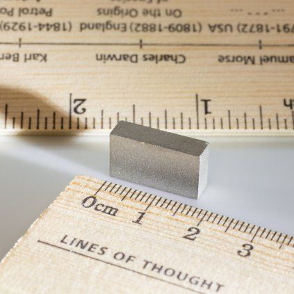
Samarium block magnet 18 × 10 × 5 mm
An SmCo magnet suitable for technical applications with higher operating temperatures and a need for good corrosion resistance.
- Dimensions 18 × 10 × 5 mm
- SmCo material
- Temperature resistance up to 300 °C
- Without a conventional protective surface coating
- Product code 35054
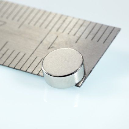
Neodymium cylinder magnet 6 × 2.5 mm
A small neodymium magnet with increased temperature resistance and poles on opposite sides of the cylindrical surface.
- Diameter 6 mm
- Height 2.5 mm
- Weight 0.53 g
- Magnetic grade N35H
- Diametrical magnetisation
- Nickel-plated surface
- Temperature resistance up to 120 °C
- Product code 20472
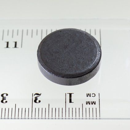
Ferrite cylinder magnet 20 × 5 mm
An affordable ferrite magnet for simple holders, demonstrations and less demanding technical projects.
- Diameter 20 mm
- Height 5 mm
- Pull force 2.2 N
- Weight 7.15 g
- MF8T isotropic ferrite
- Temperature resistance up to 200 °C
- Product code 10407
Frequently asked questions
Does MRI use X-rays?
No. It uses magnetic fields and radiofrequency energy rather than ionising X-rays.
Why does MRI use hydrogen?
Its protons are very abundant in the body, especially in water and fat, and provide an easily measured signal.
What causes the scanner’s loud noises?
Rapid switching of the gradient coils in a strong magnetic field causes mechanical vibrations.
Is MRI suitable for everyone?
Not automatically. Implants, metal objects in the body and other health factors must be assessed before the examination.
Is MRI the same as CT?
No. CT uses X-rays, while MRI works with a magnetic field and radiofrequency pulses.
Summary
MRI uses hydrogen protons in the body. A strong field creates net magnetisation, a radiofrequency pulse tips it, and a signal is produced as the protons return to equilibrium.
Gradient fields add positional information, and a computer builds a detailed image from the measured signals. MRI does not use ionising radiation but requires thorough screening for metal objects and implants.
Discover different types of permanent magnets
Compare samarium, neodymium and ferrite magnets by size, magnetisation and temperature resistance.